IMAGING
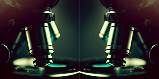
Advanced optical imaging technologies are essential tools for the context analysis at the tissue and animal scale. SCALE-UP is expanding the portfolio of instruments already present in the Department (single-photon confocal microscope, several fluorescence and bright field microscopes…) with:
– a new confocal system with laser scanning head, also equipped with light sheet, and with an automatic tracking system.
The new microscope enables analysis on different scales, ranging from reconstituted in vitro biological systems to high-resolution spatial and temporal analysis of samples with complex three-dimensional structures (bio-synthetic materials, organoids, zebrafish embryos and larvae, murine tissues, human tissues). The use of fluorescent probes allows for the integration of molecular and structural analysis with functional data. This setup also allows for the examination of tissue and organ samples subjected to clarification and expansion microscopy processes.
– the Ramona optics MCAM imaging system, a scalable multi-camera array microscope for rapid and high-throughput screening analyses in brightfield and fluorescence settings. By collecting data from up to 96 cameras, MCAM computationally generates gigapixel-scale images and movies.